|
||
|
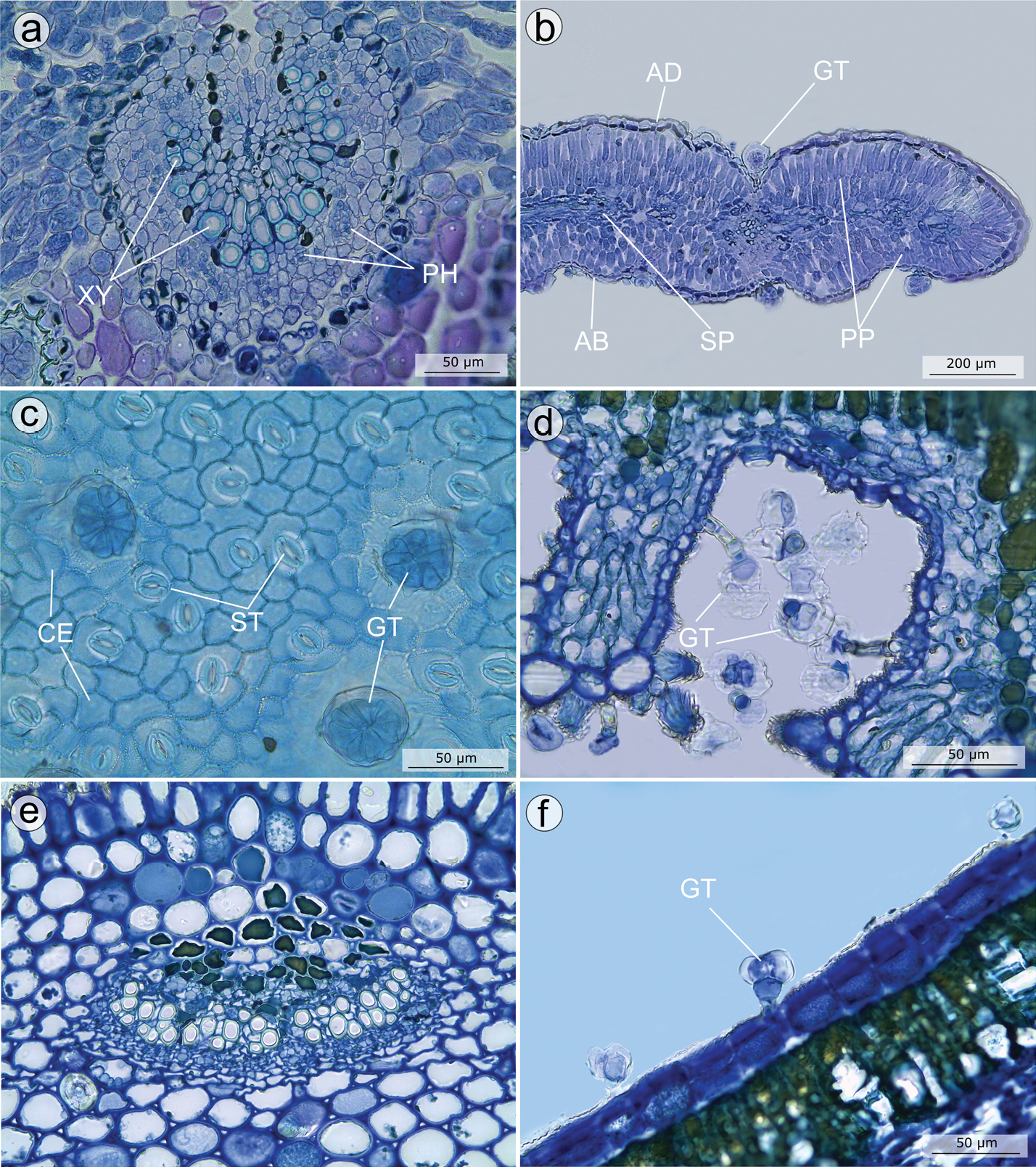
Anatomical features of leaves in the Trembleya s.s. clade of Microlicia A–C Microlicia altoparaisensis: A cross section of the mid-vein, showing an amphicribral arch-shaped vascular bundle B cross section of the leaf, showing the isobilateral mesophyll, the wavy epidermis and a glandular trichome in a depression C Adaxial surface of the epidermis in frontal view, with stomata and glandular trichomes D, E Microlicia chamissoana: D leaf stomatal crypt in cross section, with glandular trichomes E petiole in cross section, showing an amphicribral vascular bundle F Microlicia flaviflora, cross section of the leaf blade showing a reduced glandular trichome. AB: Abaxial surface; AD: Adaxial surface; CE: Common epidermal cells; GT: Glandular trichome; PP: Palisade parenchyma; SP: Spongy parenchyma; ST: Stomata. All photos by A.A.O. Carmo. Voucher specimens: A–C Pacifico & Bressan 380 (CAS, HUEM, SPF) D, E Pacifico & Carmo 154 (HUEM, UEC) F Mello-Silva et al. 509 (SPF). |