|
||
|
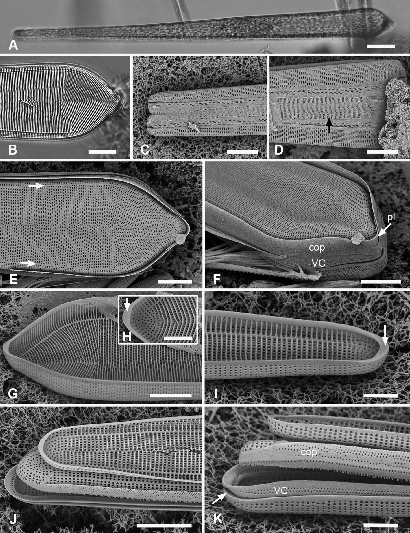
Synedrosphenia gomphonema A live specimen in valve view showing heteropolar cell shape and plastids (GU44BJ-4) B apical portion of cleaned valve in LM showing change in stria orientation where cell narrows (GU44BD-4) C, D recently divided frustule in girdle view with details of basal and apical poles, respectively, showing heteropolarity; black arrow indicates slit formation on the copula (GU44BJ-4) E, F valvar view of frustule at 0° and 60° tilt, respectively, arrows on E showing location of annulus, labels on F naming the girdle bands (abbreviations as before) G–I apical and basal portions of valve, internal view tilted 60°, showing pseudosepta at poles (H, I arrows) and transverse and longitudinal costae (TK4); inset (H) shows tip of same valve at 0° tilt with lack of costae between the last few striae but continuation of annulus J, K basal portions of a broken, recently divided frustule from Chuuk, viewed at 0° and 60° tilt, respectively, valves and girdle bands of one daughter cell with one valve of other daughter on top; arrow shows the notch between valve and valvocopula (TK4). Scale bars: 25 µm (A); 10 µm (B–G, I); 5 µm (H, J). |