|
||
|
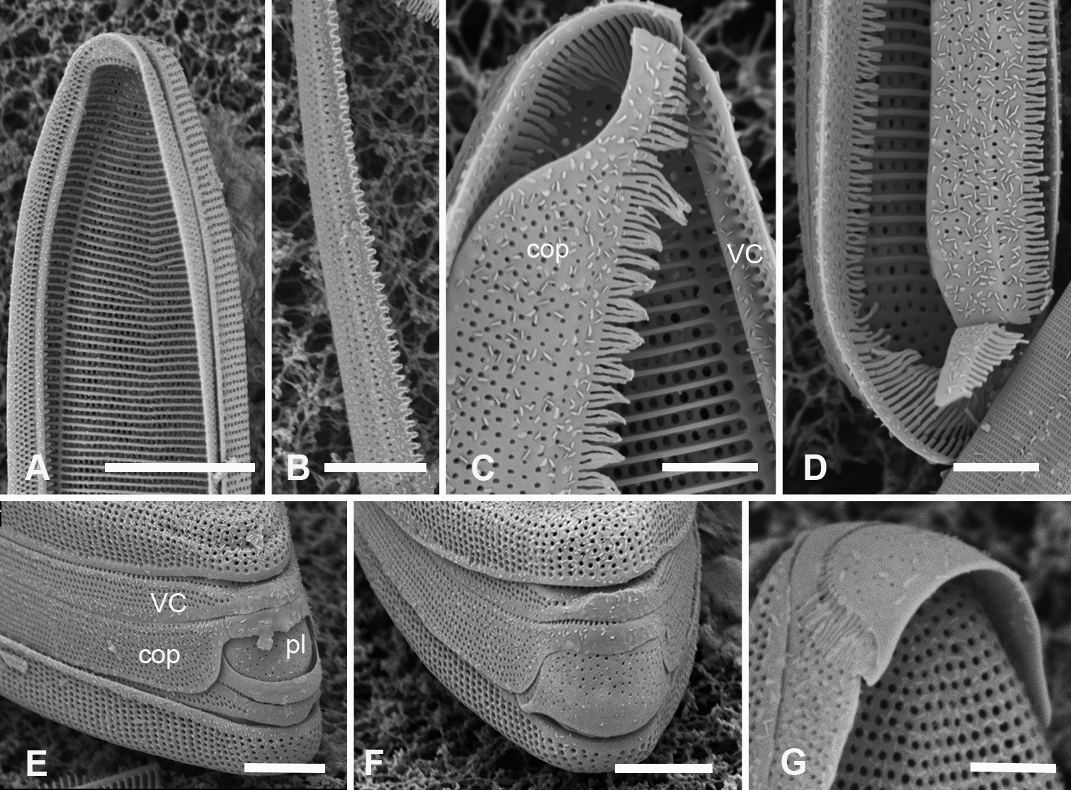
Synedrosphenia parva sp. nov., cont. All GU75A-4 A, B valvocopula showing apical internal side from abvalvar position, and portion of middle from advalvar side, respectively, showing row of pores along base of pars interior and fimbriae C internal view of apical pole with valvocopula in situ and the copula, broken in the middle, covering the valvocopula around the apical pole and left side D basal pole of same specimen, again showing the long fimbriae overlapping the valvocopula E, F frustules tilted to show polar (apical) architecture of, respectively a recently divided cell, where the pleura is in place between the copulae of the two cingula, and a non-dividing cell G detail of a pleura loosened from its position and showing some of the fimbriae (still tucked under the copula at top of image). Scale bars: 10 µm (A); 5 µm (B, D–F); 2.5 µm (C, D, G). |