|
||
|
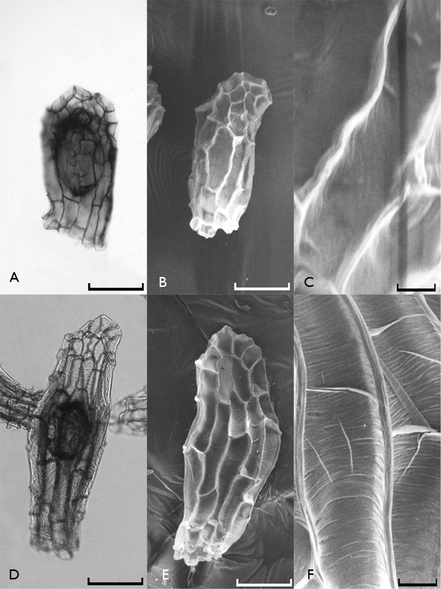
Light microscope (A, D) and scanning electron microscope (B, C, E, F) photographs of Orchis simia subsp. simia (A, B, C) and Neotinea tridentata subsp. tridentata (D, E, F) seeds. Scale bars: 0.1 mm (A, B, D, E) and 0.01 mm (C, F). |
|
||||||||
| Part of: Güler N (2016) Seed micromorphology of Orchis Tourn. ex L. (Orchidaceae) and allied genera growing in Edirne province, Turkey. PhytoKeys 68: 9-25. https://doi.org/10.3897/phytokeys.68.8746 |