|
||
|
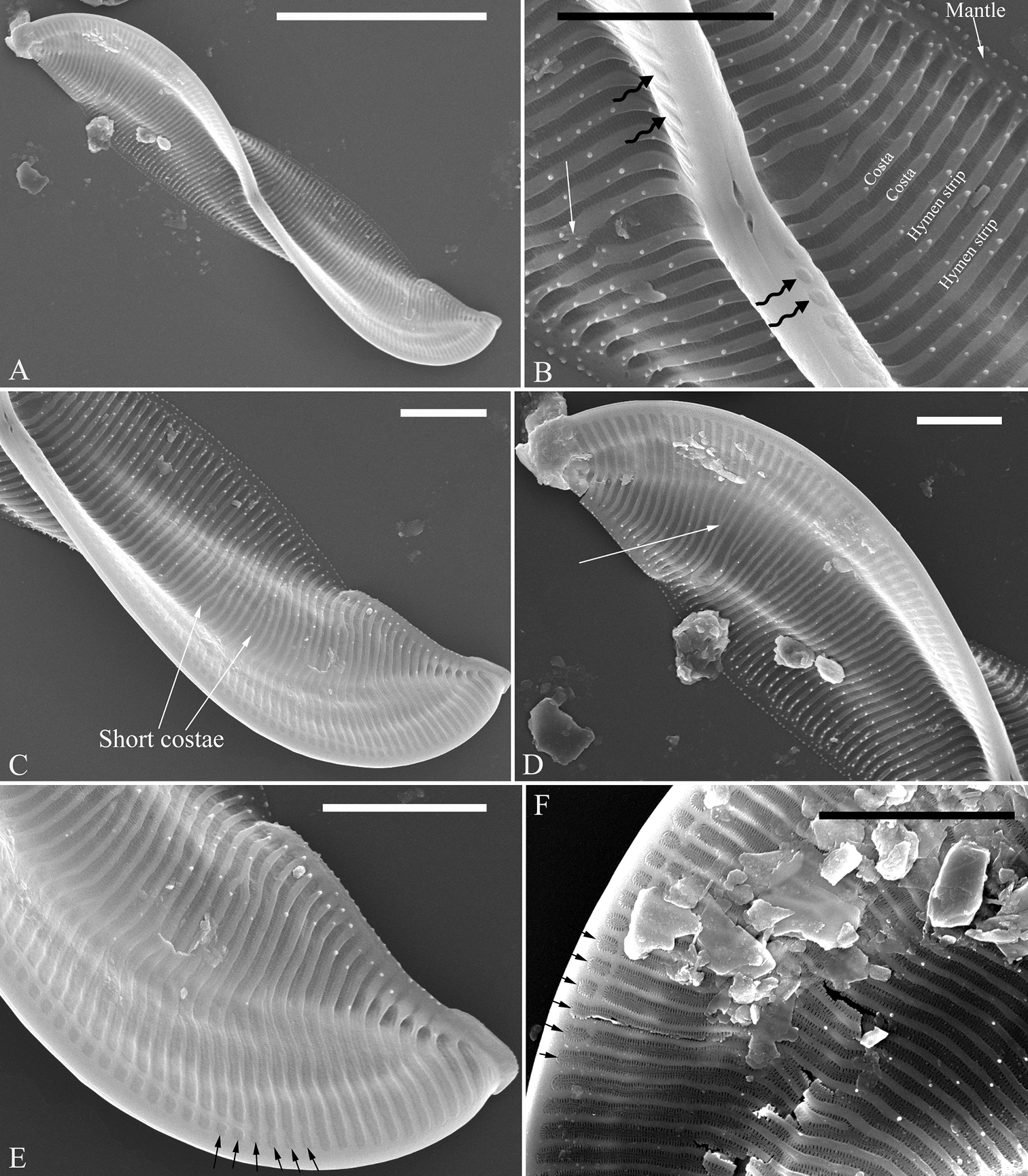
A–F Entomoneis sinensis sp. nov., valve external view, SEM A one whole valve showing the Ƨ-shaped keel outline B central part from Fig. A note hymen strips, costae, mantle, warts, forked costa (arrow) and one separated row of rounded areolae at each side of the raphe (wavy arrows) C, D two apices from Fig. A note the short costae (two arrows in Fig. C) and two costae merging into one (arrow in Fig. D) E, F details showing one separated row of rounded areolae terminating before the apex (six arrows, respectively). Scale bars: 10 μm (A); 2 μm (B–F). |