|
||
|
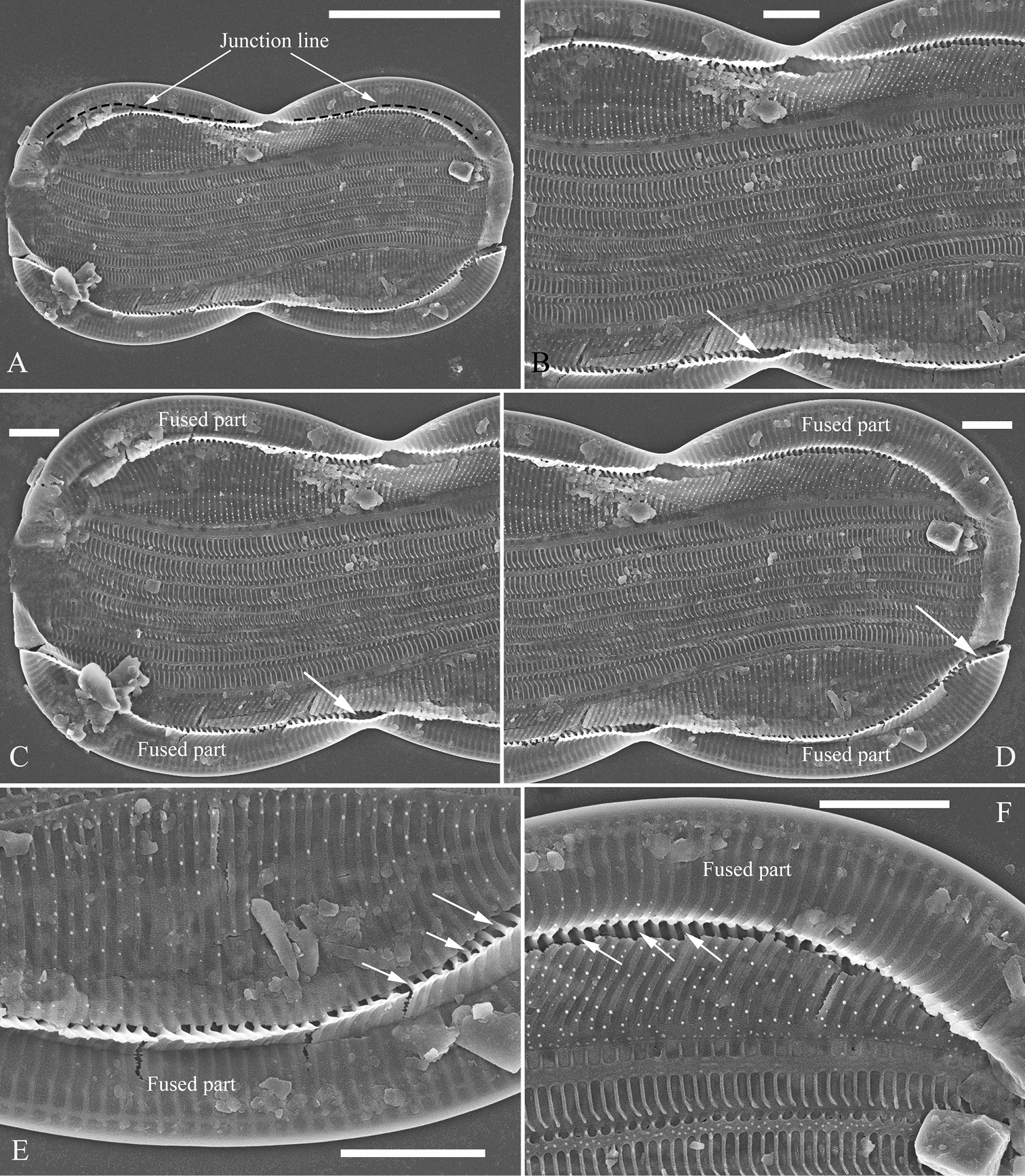
A–F Entomoneis sinensis sp. nov., girdle view, SEM A one broken frustule, note the simple arcuate junction lines B–D details from Fig. A note the fused parts of two sides of the keel and the subraphe canal connecting the cell lumen only near the central (Figs B and C, arrow, respectively) and the distal raphe ending (Fig. D, arrow) E, F details from Fig. A note the short, bar-like basal fibulae (three arrows, respectively). Scale bars: 10 μm (A); 2 μm (B–F). |