|
||
|
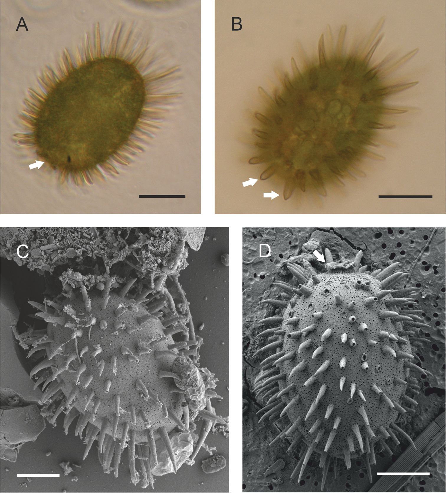
Visualisation of the morphology of Trachelomonas bituricensis var. lotharingia by optical and electron scanning microscopy. A Overall appearance of lorica under an optical microscope, with a short collar with undulated edge (marked with arrow) B Detailed image of the lorica surface with three types of spines: long, curved spines at the antapical part of the lorica; shorter spines on the lorica’s body; and several straight spines near the apical pore (marked with arrows). Note the numerous disc-like chloroplasts in the cell C, D Loricas viewed by scanning electron microscopy, with clearly visible punctuation at the surface, and short collar with undulated edge (marked with arrow). Scale bars = 10 μm. |