|
||
|
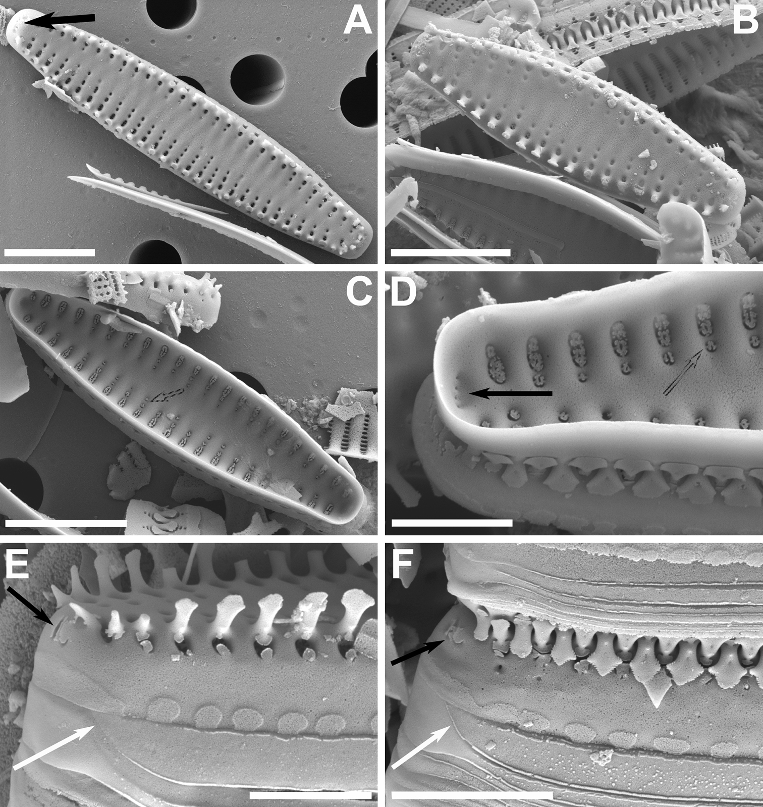
A–F SEM images of Pseudostaurosira occulta sp. nov. A, B external views of valves showing apical pore fields (black arrow in A) and other features C internal view of valve showing depressions containing the striae (dashed arrow) D internal close-up showing apical pore field depression (black arrow) and striae depression (dashed line) E close-up on valve apex showing flaps on apical pore field (black arrow) and open girdle element (white arrow) F close-up on cell-cell connection showing apical pore field covered with flaps (black arrow) and open girdle element (white arrow). Scale bars: 2 µm (D, E); 3 µm (F); 5 µm (A–C). |