|
||
|
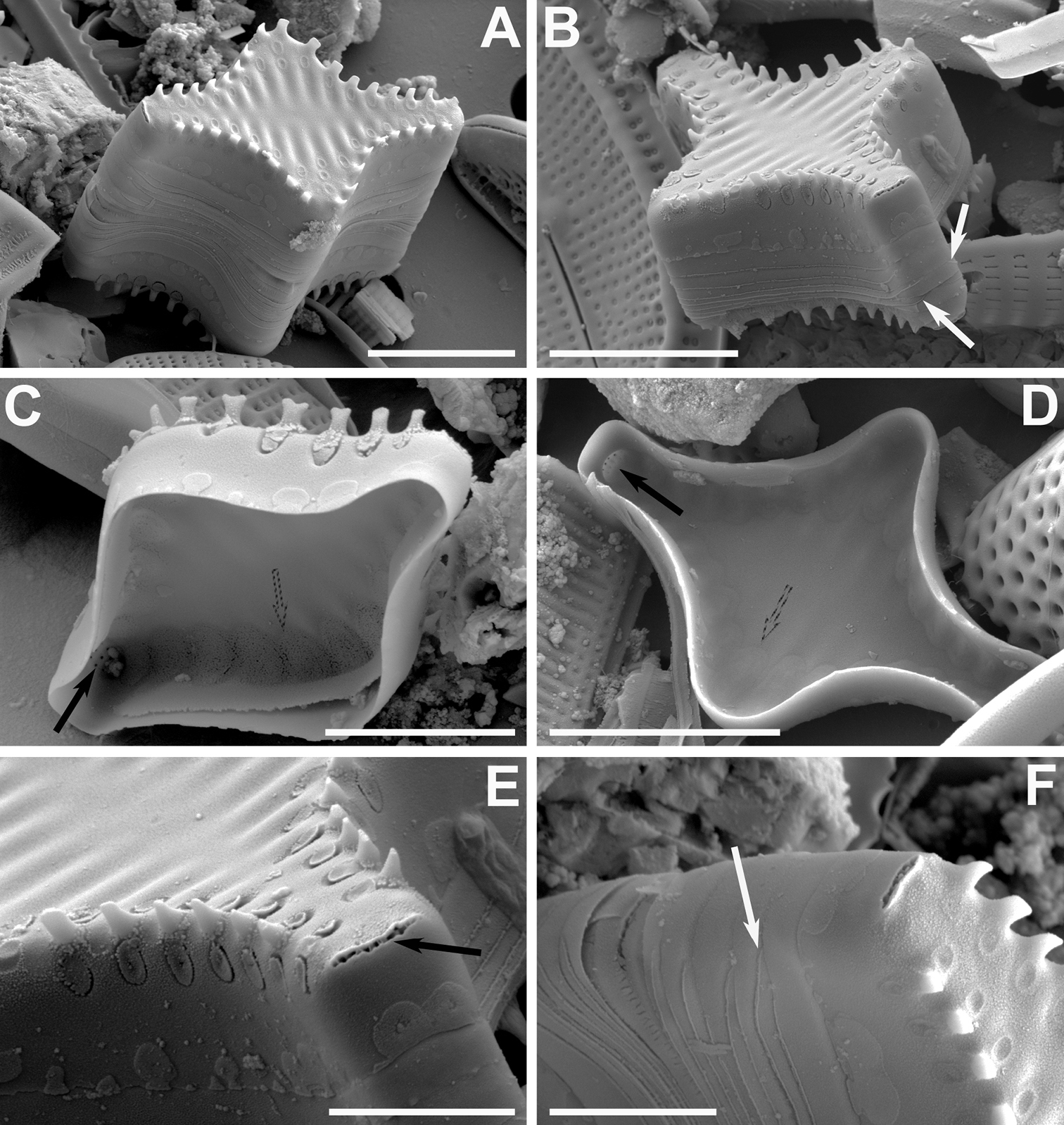
A–F SEM images of Pseudostaurosira frankenae sp. nov. A, B titled frustules showing external features; notice open girdle elements in B (white arrows) C, D internal view of valves showing internal elliptic depositions on striae (dashed arrows) and depression of the apical pore field (black arrows) E close-up of frustule apex (B) showing the externally occluded apical pore field, showing only a single row of poroids (black arrow) F close-up of frustule tip (A) showing open girdle bands (white arrow). Scale bars: 2 µm (E, F); 3 µm (C); 5 µm (A, B, D). |