|
||
|
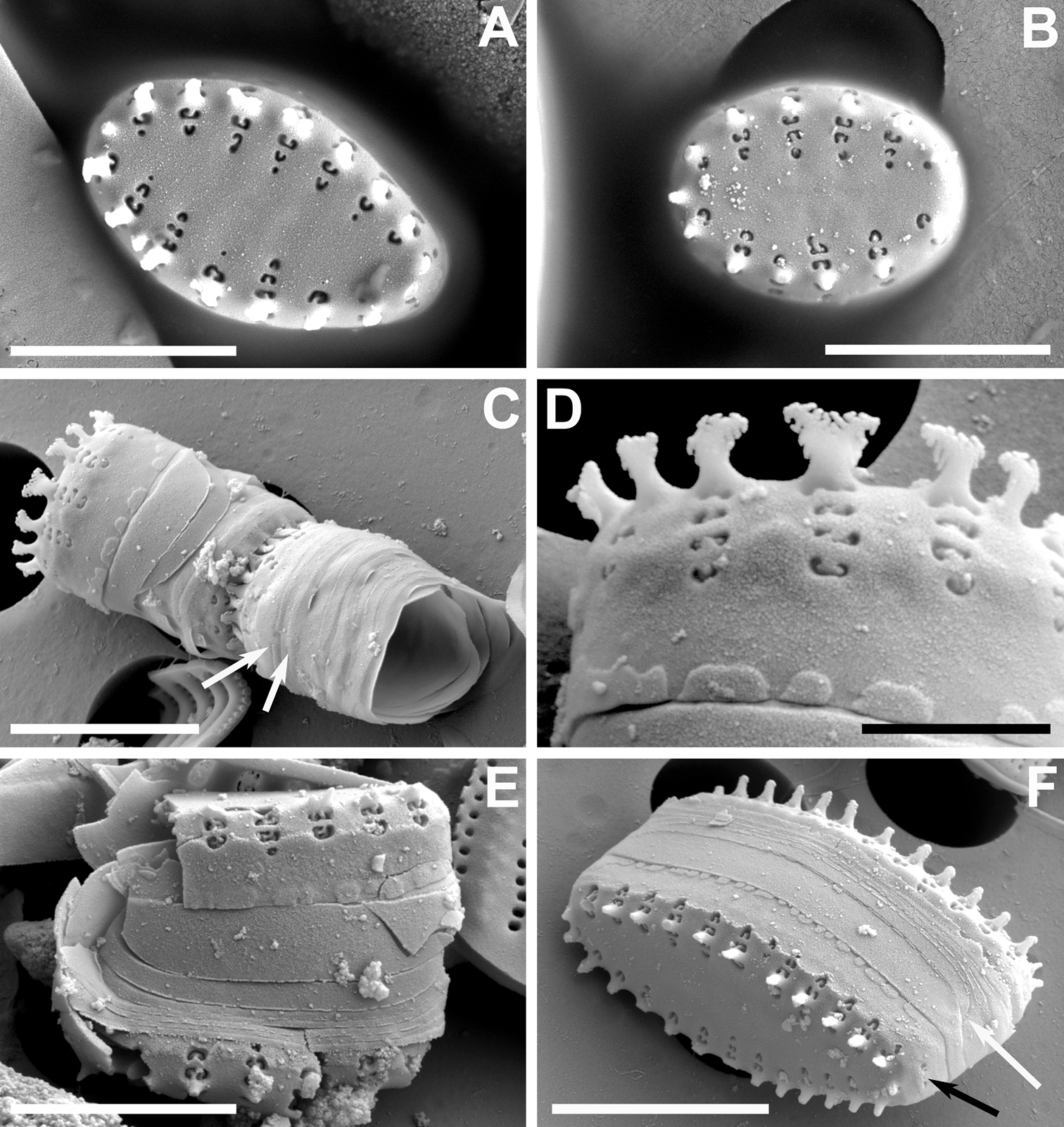
A–F SEM images of Pseudostaurosira heteropolaris sp. nov. from the Bolivian Altiplano A, B Valve views showing striation pattern and features of the axial area, virgae, vimines, and spine location C side view of complete frustule and neighboring cell showing girdle bands (white arrows denote open copulae). White arrows point to open girdle elements D close up of C showing details of spines with bifurcations with pinnatifid projections, characteristics of the striae on valve mantle and the features of the blisters E broken frustule with girdle bands. Pattern of volae within areolae is also shown F frustule in side, tilted view. Notice open copulae (white arrow) and reduced apical pore field with cavernous poroids (black arrow). Scale bars: 1 µm (D); 3 µm (A–C, E); 4 µm (F). |