|
||
|
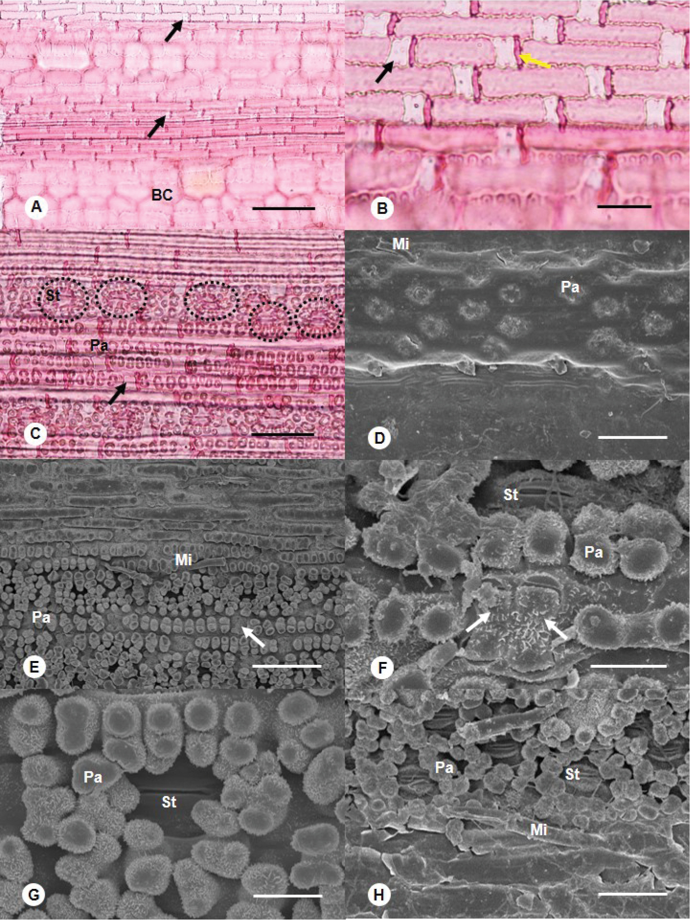
Leaf micromorphology of Buergersiochloa bambusoides A–C leaf surface observed under LM A adaxial surface showing long cells, bulliform cells with sinuous anticlinal walls and saddle-shaped silica bodies B detail of saddle-shaped silica bodies and cork cells C abaxial surface showing silica bodies, abundant papillae and papillae encircling the stomata (dotted circles) D–H leaf surface observed under SEM D adaxial surface with very small papillae and the basal cell of a broken bicellular microhair E abaxial surface showing microhairs, abundant papillae on costal and stomatal cell rows, and long cell papillae encircling the stomata F detail of two adjacent saddle-shaped silica bodies, papillae and stoma on the abaxial surface G detail of papillae encircling a stoma H detail of panicoid type bicellular microhairs. BC: Bulliform cell; Mi: bicellular microhair; Pa: papilla; St: stoma. Black or white arrows: silica body; yellow arrow: cork cell. Scale bars: 100 µm (A, C); 25 µm (B, H); 10 µm (D, G); 50 µm (E, F) |