|
||
|
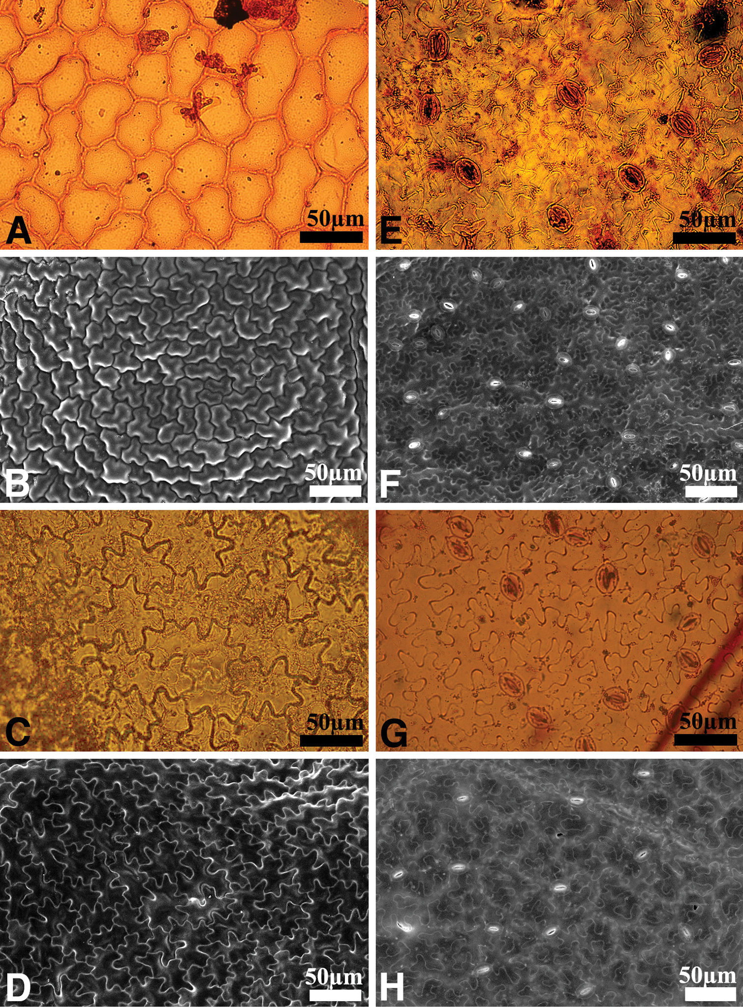
Differences in the leaf epidermis of Astilbe uljinensis and Astilbe chinensis A, B, E, F A. uljinensis C, D, G, H A. chinensis; Left: adaxial surface; Right: abaxial surface A, C, E, G Light Microscope image B, D, F, H by Scanning Electron Microscope image. Scale bars: 50 µm. |