|
||
|
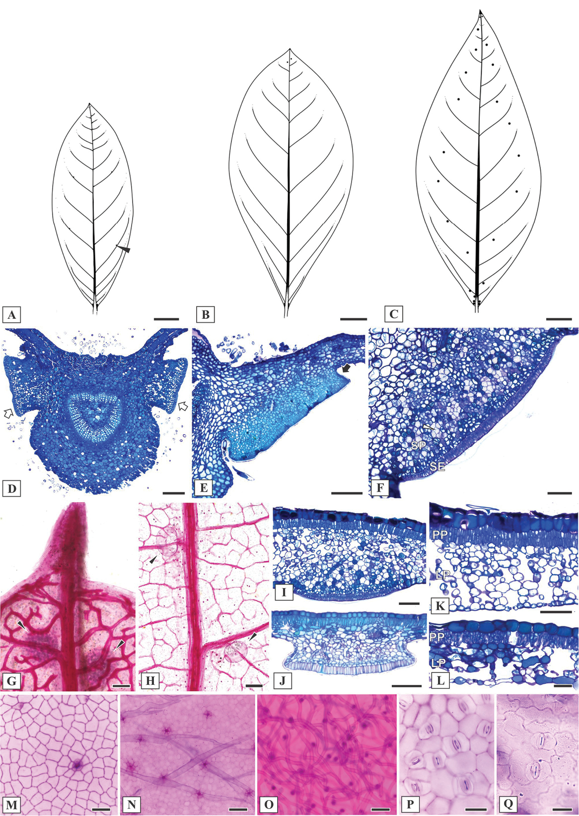
Leaf morphoanatomy of Mcvaughia species. A patterns of leaf glands distribution on the abaxial leaf surface of M. bahiana B patterns of leaf glands distribution on the abaxial leaf surface of M. piauhiensis C patterns of leaf glands distribution on the abaxial leaf surface of M. sergipana D transverse section of leaf base showing the basilaminar pair of stalked glands (white arrows) E basilaminar leaf gland with a stalk (black arrow) in M. piauhiensis F basilaminar gland in M. sergipana showing a sessile position (SE= anatomical arrangement with secretory epidermis, SP= vascularized secretory parenchyma) G–H laminar glands on the apex of cleared leaves of M. sergipana and M. bahiana respectively, note the apical tooth (G) I sessile laminar glands in M. sergipana J stalked laminar gland in M. piauhiensis K–L transverse sections of the leaf blade; mesophyll with uniserial palisade-like parenchyma and spongy parenchyma composed by several or few layers in M. sergipana and M. bahiana, respectively; note the idioblast with druse crystals at the mesophyll (white arrow) and the stomata distribution at the abaxial leaf surface (black arrow) M–N adaxial epidermis surface of M. piauhiensis and M. sergipana, showing scars of malpighiaceous trichomes O abaxial epidermis surface of trichomes abundance in M. bahiana P–Q outline of the anticlinal epidermal cell walls: straight in M. sergipana (P) and sinuous in M. bahiana (Q). Laminar scale bars: 1 cm (A–C), 100 μm (D, F–K, N–O), 150 μm (E), 50 μm (L–M, P–Q). |