|
||
|
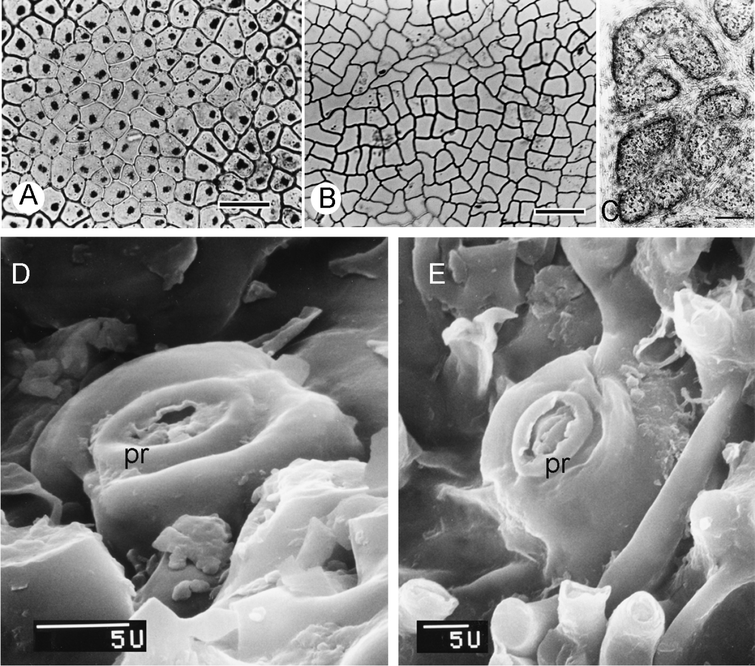
LM photograph and SEM micrographs of leaf adaxial, abaxial surface and stomata in Curarea: A isodiametric epidermal cells, C. candicans (Jansen-Jacobs et al. nr-1995) B triangular to hexagonal epidermal cells, C. cuatrecasasii (Kernan & Phillips 1147) C abaxial surface showing areoles (black dotted) in C. toxicofera (van der Werff & Vásquez 13990) D, ESEM micrographs of projecting stomata with distinct peristomatal rim D C. tecunarum (Ortiz et al. 143) E C. tomentocarpa (Reynel & Meneses 5025). Scale bar: 50 µm (A); 100 µm (B), 0.4 mm (C), pr = peristomatal rim. |