|
||
|
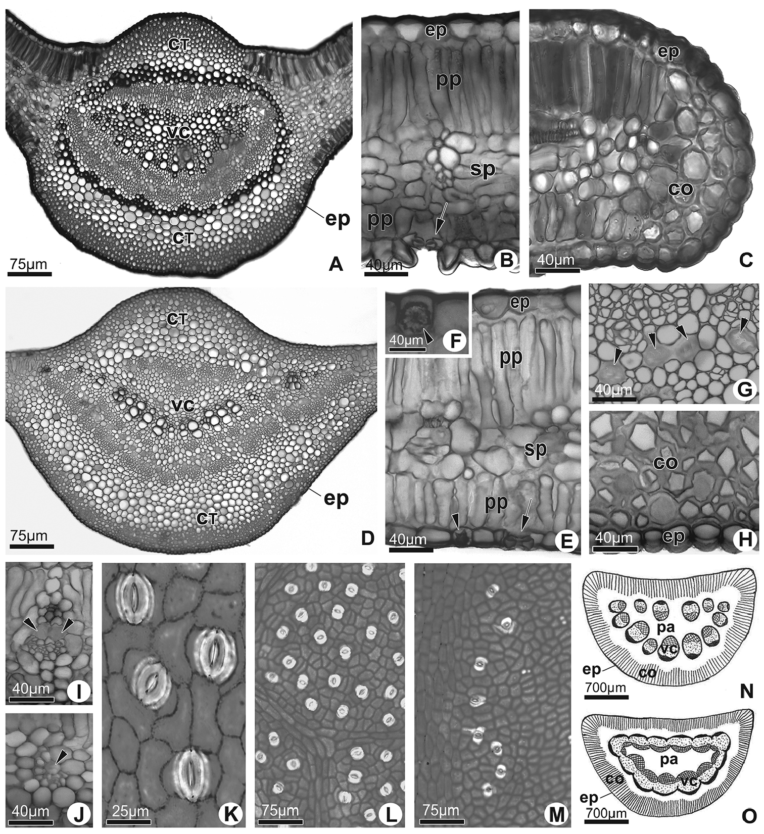
Leaf anatomy of species. A–J Cross section of the leaf blade. Manihot fallax (A–C): A Median portion of the midvein B Median portion of the leaf blade; abaxial epidermis papilose and stomata indicated by arrow C Edge. Manihot attenuata (D–J): D Median portion of the midvein E Median portion of the leaf blade; note stomata (arrow) and epidermal idioblast with druse (arrowhead) F Detail of epidermal idioblast with druse (arrowhead) G Laticifers in the midvein phloem (arrowheads) H Detail of the epidermis and collenchyma in the midvein I Vascular bundles with parenchymatical sheath in the median portion of the leaf blade (arrowheads correspond to laticiferous) J Vascular bundles in the median portion of the leaf blade (arrowheads correspond to laticifers) K–M Dissociated epidermis of both species. Manihot fallax. K. Paracytic stomata. Manihot attenuata (L–M): L Stomata distribution in the abaxial surface of the leaf blade M Stomata distribution in the adaxial surface of the leaf blade lateral to midvein N and O Schematic representation of the petiole in cross section N Manihot fallax O Manihot attenuata. CT: cortex; VC: vascular cylinder; co: collenchyma; ep: epidermis; pp: palisade parenchyma; sp: spongy parenchyma; pa: medullar parenchymatic. |